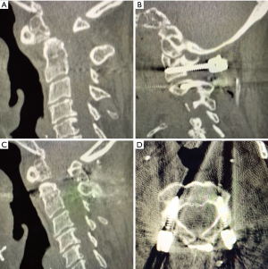
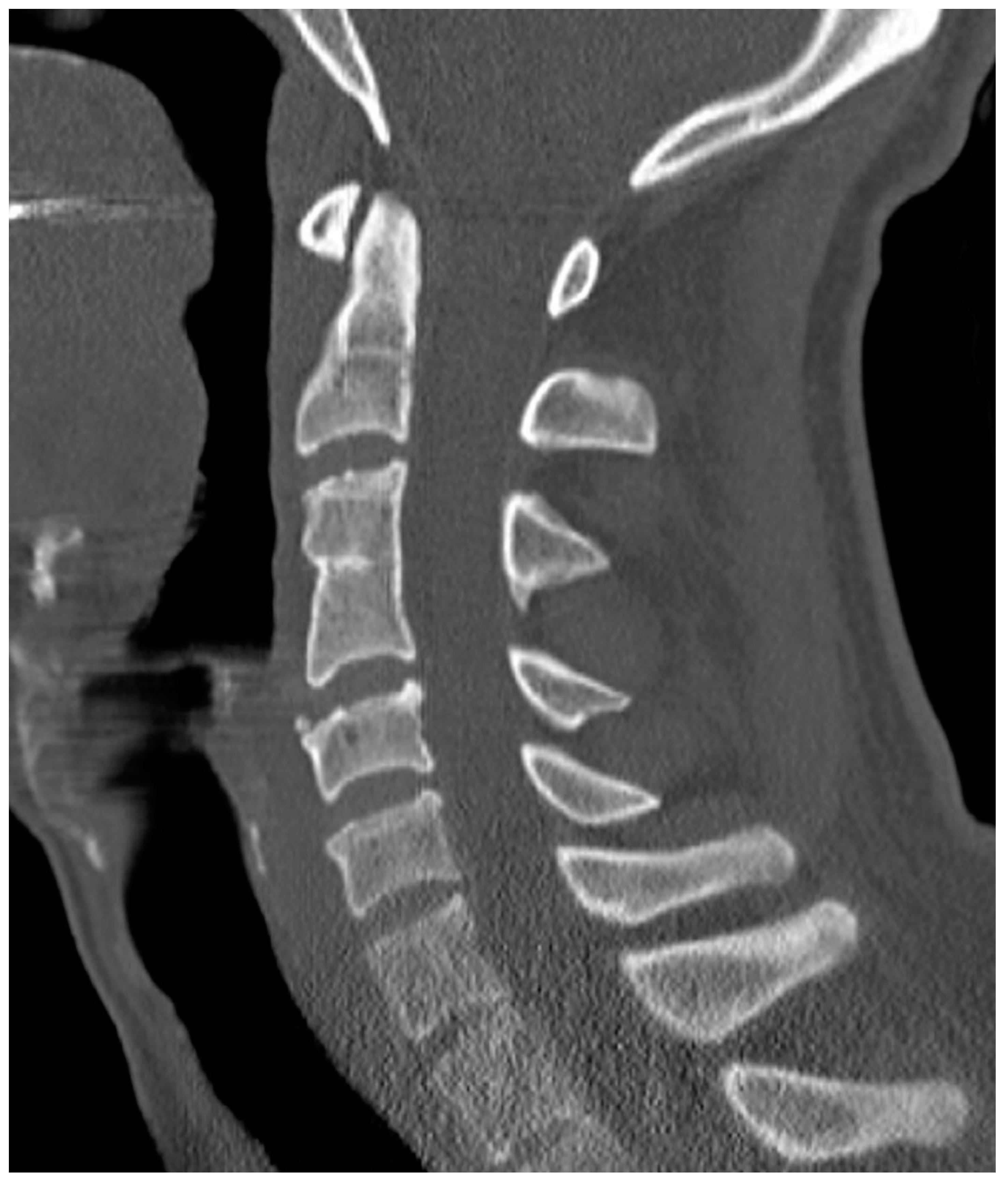
CT image of C2–3 congenital fusion. A Vertebral body fusion and lamina
By A Mystery Man Writer
Description
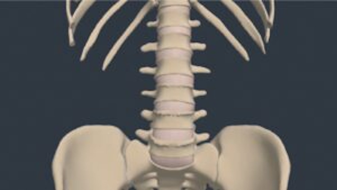
Lumbar Vertebrae - Physiopedia

CT image of C2–3 congenital fusion. A Vertebral body fusion and lamina

Surgical Management of Complex Spinal Deformity
Posterior atlantoaxial fusion: a comprehensive review of surgical techniques and relevant vascular anomalies - Chen - Journal of Spine Surgery

Anatomical analysis of the C2 pedicle in patients with basilar invagination
C1-C2 Facet Joint Penetration by C2 Pedicle Screws: Influence of Local Anatomy, Bone Mineral Density, and Screw Length

Anatomical analysis of the C2 pedicle in patients with basilar invagination
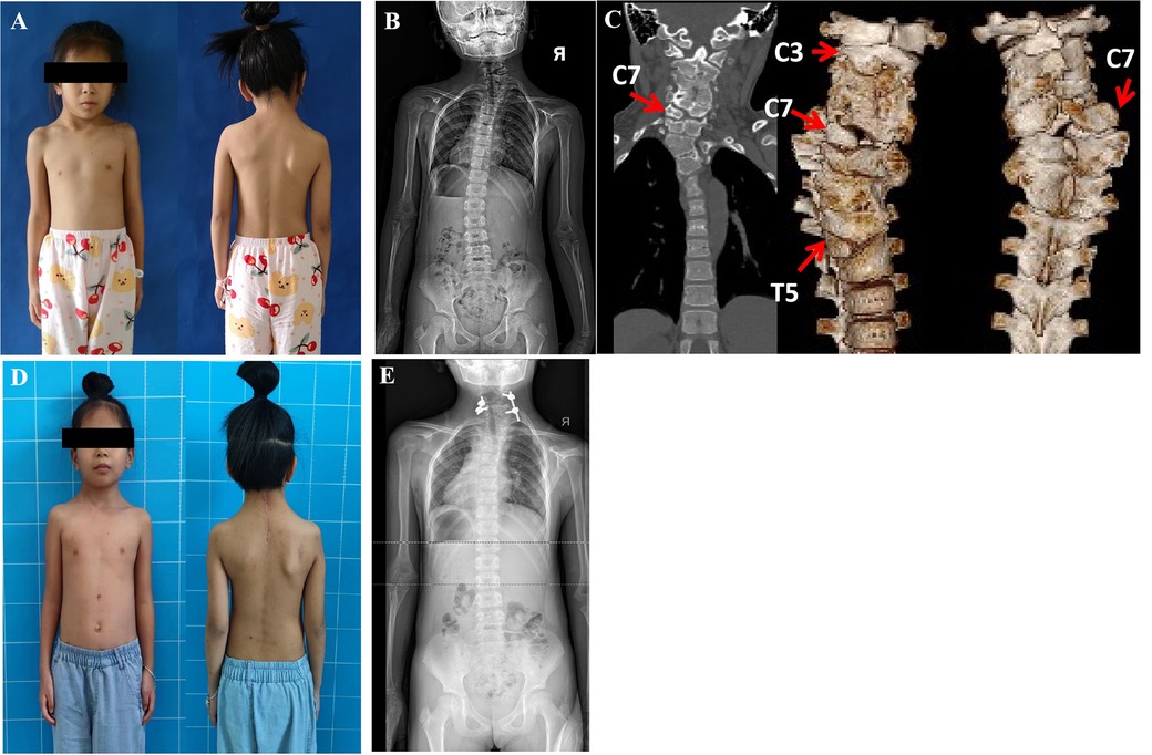
Frontiers Minimally invasive spine surgery strategy for congenital cervicothoracic scoliosis in children: Less blood loss and shortened segmental fusions/fewer pedical screws

SCIWORA in patient with congenital block vertebra

Xiangyang Ma's research works Wuhan General Hospital of Guangzhou Military Command, Wuhan and other places

Anatomical analysis of the C2 pedicle in patients with basilar invagination

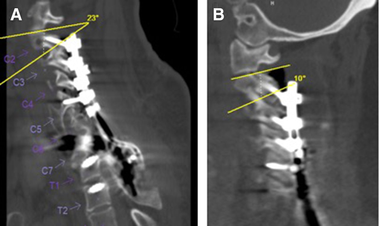
CT scan of the cervical spine, a the sagittal image demonstrates fusion

Cervical Radiculopathy - Spine - Orthobullets
Diagnostics, Free Full-Text
from
per adult (price varies by group size)