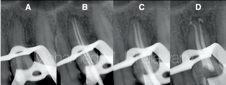
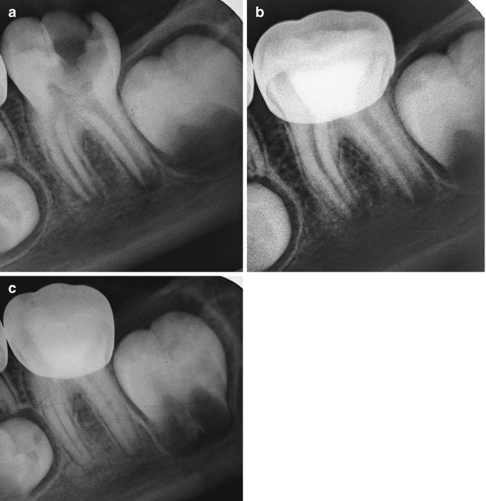
A) Preoperative intraoral periapical (IOPA) radiograph of 36. B) Post operative (IOPA) radiograph of 36. C) 1 month follow up IOPA radiograph of 36. D) 6 months follow up IOPA radiograph of
By A Mystery Man Writer
Description
A) Preoperative intraoral periapical (IOPA) radiograph of 36. B) Post operative (IOPA) radiograph of 36. C) 1 month follow up IOPA radiograph of 36. D) 6 months follow up IOPA radiograph of 36. E) 1 year follow up IOPA radiograph of 36. - IP Indian J Conserv Endod - clinical and preclinical conservative /restorative de

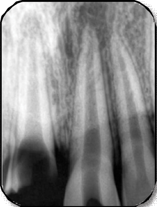
a) Preoperative IOPA radiograph of tooth #36. (b) Intraoral image
Pre-operative X-ray: suggested or obligatory - Style Italiano Endodontics

A) Preoperative intraoral periapical (IOPA) radiograph of 36. B) Post

Nonsurgical Management of Periapical Lesion: A Case Series

JaypeeDigital
jcdr-11-ZD05-g006.jpg
jcdr-11-ZD05-g003.jpg

A) Preoperative intraoral periapical (IOPA) radiograph of 36. B) Post

Radiograph sem
Strategies for Pulp Therapy in Immature Permanent Teeth

File:Intraoral Periapical Radiograph (IOPA) showing Deciduous(Milky or Primary) Tooth 75 and developing crown of Permanent or Secondary Teeth 35, 36 and 37.jpg - Wikipedia
PDF) Direct pulp capping with bioactive materials – A case series

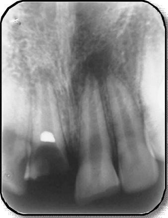
Preoperative and postoperative radiographs of 14 and 24 showing three

JaypeeDigital
from
per adult (price varies by group size)