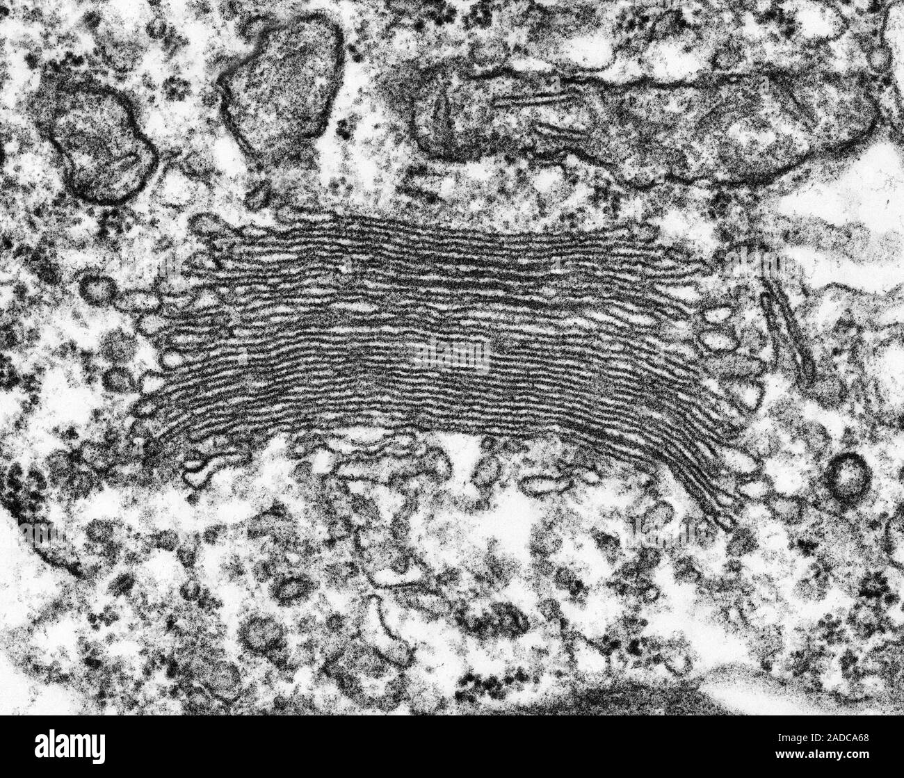
Under high magnification, it shows the stacks of Golgi apparatus
By A Mystery Man Writer
Description

Do Now 9/9 The diagram shows a stage micrometer viewed with an eyepiece graticule scale, using a magnification of ×400. Using the same magnification, a. - ppt video online download
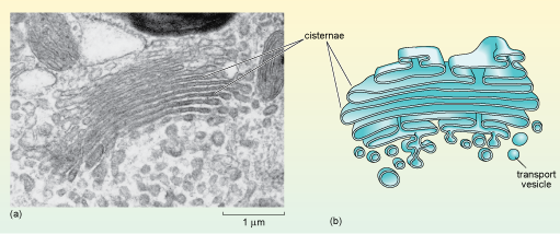
A tour of the cell: 4.7 The Golgi apparatus

In vivo characterization of Drosophila golgins reveals redundancy and plasticity of vesicle capture at the Golgi apparatus - ScienceDirect

Non-canonical features of the Golgi apparatus in bipolar

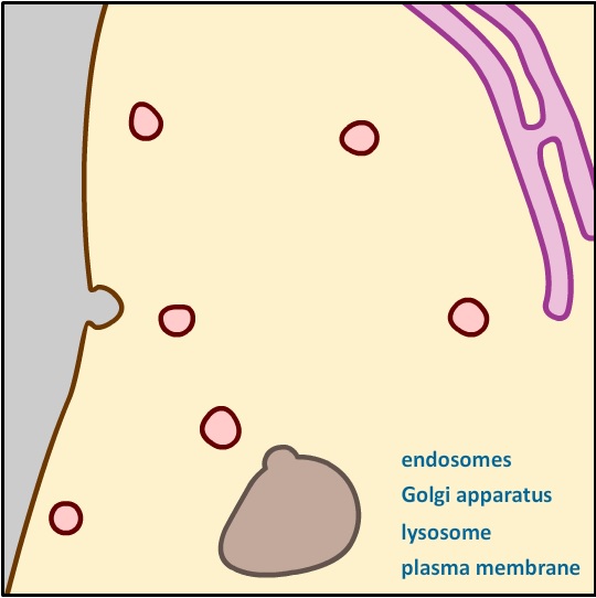
The Endomembrane System Biology for Non-Majors I
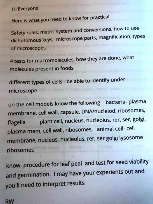
SOLVED: Hi Everyone, here is what you need to know for practical safety rules, the metric system and conversions, how to use dichotomous keys, microscope parts and magnification, types of microscopes, macromolecules

Quantifying Golgi structure using EM: combining volume-SEM and

GOLGI - Stock Photos, Illustrations and Images - Album
Scanning electron micrograph (TEM) of Golgi apparatus, stacks of cisternae and vesicles (Euglena sp.). The Golgi apparatus is a cell organelle in all Stock Photo - Alamy
Cell Component
from
per adult (price varies by group size)


:max_bytes(150000):strip_icc()/Bloodpressure-5204833-final-7927123aab224096bc5494908bbdc873.jpg)